Question 1:
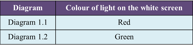
Diagram 1.1 and Diagram 1.2 show an experiment to study the effect of coloured filter on white light.
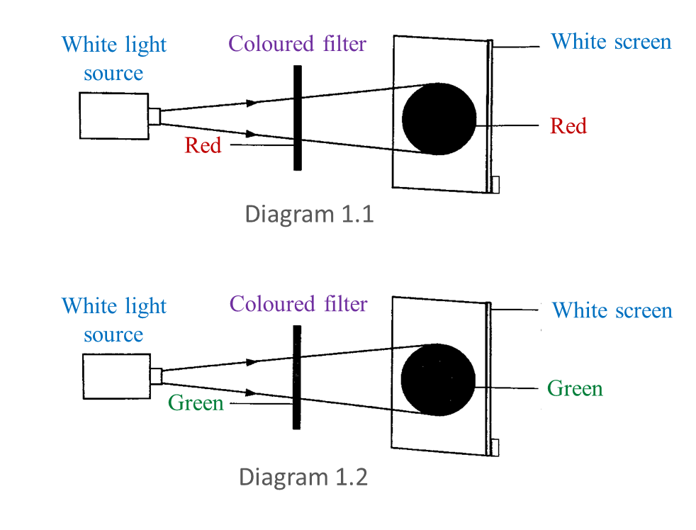
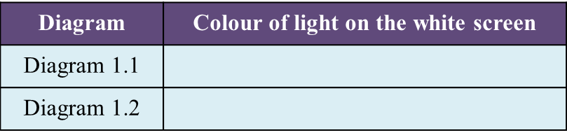
(a) Based on this experiment, state the colour of light observed on the white screen. Write down your answer in Table below. [2 marks]
(b) Write down
one hypothesis for this experiment
. [1 mark]
(c) State
one manipulated variable in this experiment. [1 mark]
(d) Predict the colour of light observed on the white screen if blue filter is used. [1 mark]
Answer:
(a)
(b) The colour on the screen is the same as the colour of the filter.
(c) The type of colour filter.
(d) Blue
Question 2:
Diagram 2.1 shows three coloured lights projected on a white screen.
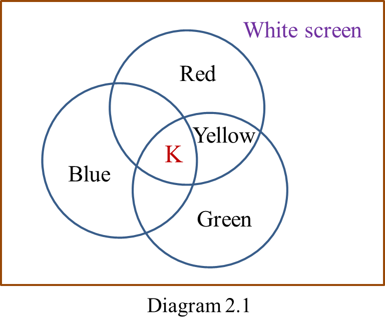
(a) Based on the coloured lights labelled in Diagram 2.1, state
(i) a primary colour, [1 mark]
(ii) secondary colour. [1 mark]
(b) What is the colour represented by K? [1 mark]
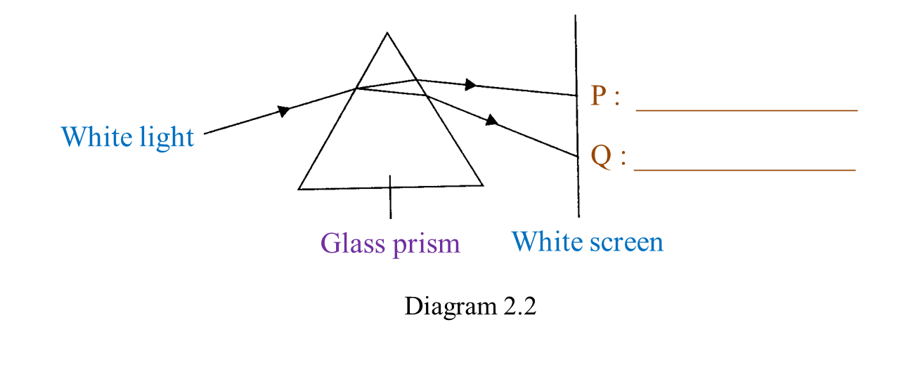
(c) Diagram 2.2 shows a white light projected through a glass prism. [2 marks]
In Diagram 2.2, write down the colours of lights P and Q.
(d) A green filter is placed as shown in Diagram 2.3.
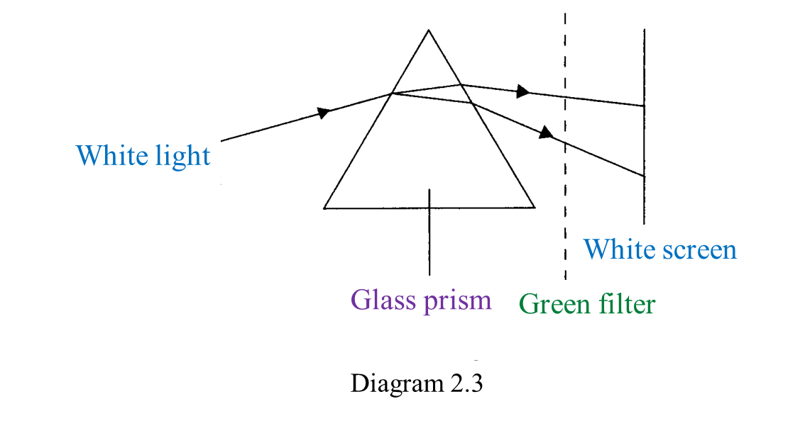
What colour is seen on the white screen? [1 mark]
Answer:
(a)(i) Blue/ Red/ Green
(a)(ii) Yellow
(b) White
(c) 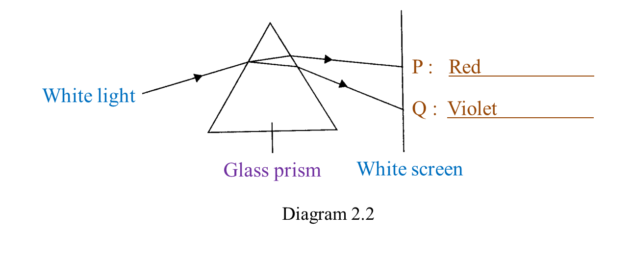 (d)
(d) Green