1.3 Blood Clotting


Hemostasis 

  1. Hemostasis is a process which causes bleeding to stop and keep blood within a damaged blood vessel. It is the first stage of wound healing.
  2. Blood clotting is an important part of hemostasis, wherein a damaged blood vessel wall is covered by a platelet and fibrin-containing clot to stop bleeding and begin repair of the damaged vessel.

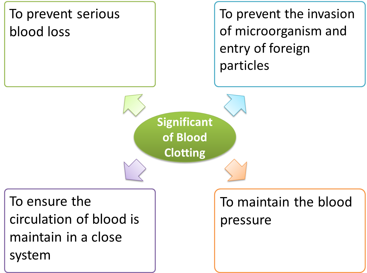
Significant of Blood Clotting


1.2.6 Regulation of Blood Pressure


  1. Pressure is how much force exerted on one unit area of a surface.
  2. Blood pressure is the pressure exerted by the blood on the walls of the blood vessels.
  3. Sometime, it is also called the arterial blood pressure as it is the pressure in the arteries.

    Normal Blood Pressure

    1. When measuring blood pressure, we measure the
      1. systolic pressure (highest pressure measured in the aorta and the arteries during ventricular contraction)
      2. diastolic pressure (lowest pressure measured after the contraction of the heart while the chambers of the heart refill with blood.)
    2. For an adult, the normal systolic pressure is 120 mm Hg whereas the diastolic pressure is 80 mm Hg.
    3. However, blood pressure may varies throughout the day.


      Factors Affecting Blood Pressure

      1. Blood pressure can be affected by the following factors:
        1. Rate of heart beat
          Higher heart rate, higher blood pressure.
        2. Volume of blood in the body
          Bigger volume, higher blood pressure.
        3. Rate and volume of blood flow
          Higher rate and volume of blood flow, higher rate of heart pumping, higher blood pressure.
        4. Resistance of blood vessels
          Higher resistance (smaller blood vessels, rough blood vessel wall), higher blood pressure. 


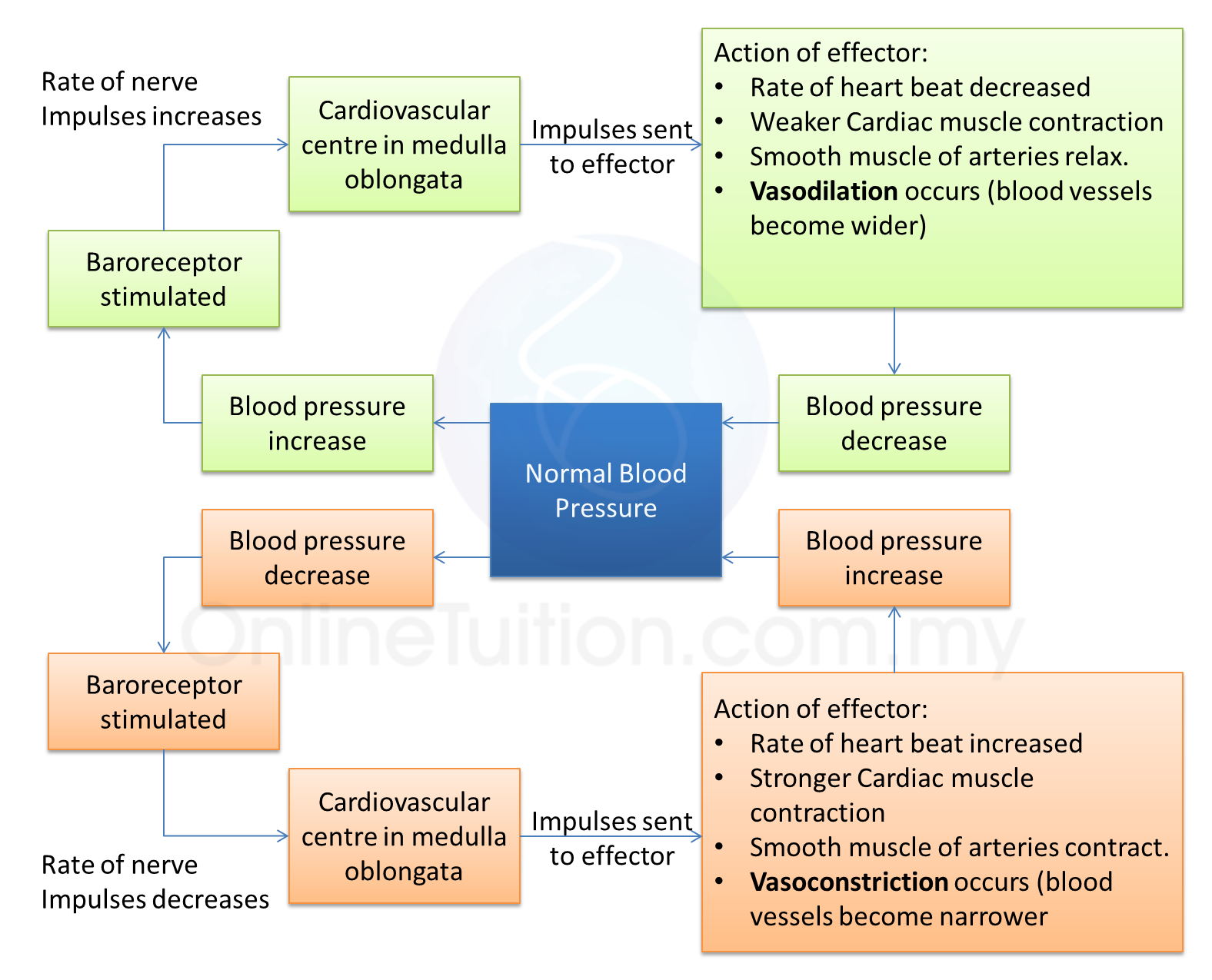
      Regulation of Blood Pressure


      Recommended Videos

      Blood Pressure Part 1

      By edunationmalaysia available under a Creative Commons Attribution  License

      Blood Pressure Part 2 (Regulation)

      By edunationmalaysia available under a Creative Commons Attribution  License

      1.2.5c Circulation of Blood in Humans

      1. The circulation of blood in human is due to the pumping of the heart.
      2. The cardiac muscles contract to produce heartbeat which pumps the blood to the whole body.

      The Cardiac Cycle

      ( Image by Madhero88 shared under the Creative Commons Attribution 3.0 Unported license. )


      1. The heartbeat is initiated by the sinoatrial node (also called the SAN or pacemaker) .
      2. The SAN consists of specialised cardiac muscle fibres that generates nerve impulses.
      3. From the SAN, the electrical impulses spread to the atrioventricular node (AVN) located at the base of the right atrium (See image above).
      4. Special muscle fibres called His Bundle and the Purkinje fibres then sends the impulses to the ventricles and then stimulates both ventricles to contract simultaneously and pumps the blood out of the heart.


      Contraction of the Skeletal Muscles Around Veins

      1. The blood pressure in the vein is low and hence make it hard for the blood to flow back to the heart.
      2. Therefore, there are skeletal muscles presence around the veins to help blood circulation.
      3. When the skeletal muscles contract, blood is forced to move along the veins.
      4. The valves in the veins prevent the back flow of the blood and hence direct the blood back to the heart.


      Recommended Videos

      How the cardiac cycle is produced by electrical impulses in the heart

      1.2.5b The Heart


      (Image by unknown author under Creative Commons Attribution-Share Alike 3.0 Unported license.)

      Structure

      1. The human heart consist of four compartment:
        1. two upper chambers (left atrium and right atrium
        2. two lower chambers (left ventricle and right ventricle)
      2. The chamber to the right is separated from the chamber to the left by a wall called septum.

      The Atria and Ventricles

      1. The atria are smaller and have thinner wall compare to the ventricles.
      2. The ventricles have thicker wall because they have to pump the blood to other organs of the body with greater pressure.
      3. Also, the wall of the left ventricle is thicker than the right ventricle because the right ventricle only has to pump blood to the lungs whereas the left ventricle has to pump blood to all parts of the body. 

      The Valves

      1. The function of the valves in the heart are to prevent the back flow of blood.
      2. There are 4 valves in human heart (as shown in figure above):
        1. the tricuspid valve
        2. the bicuspid valve
        3. the semi-lunar valve
        4. the aortic valve

      Contraction of Heart

      1. The wall of the heart is made up of cardiac muscle, which is myogenic.
      2. Myogenic contraction is the contraction of muscle without nervous stimulation.
      3. Contraction of heart is started by sinoatrial node (also known as the pacemaker)


      Recommended Videos

      How the heart works


      Cardiac Cycle


      Heart Pumping Mechanism

      By edunationmalaysia available under a Creative Commons Attribution  License

      1.2.5a The Blood Vessels

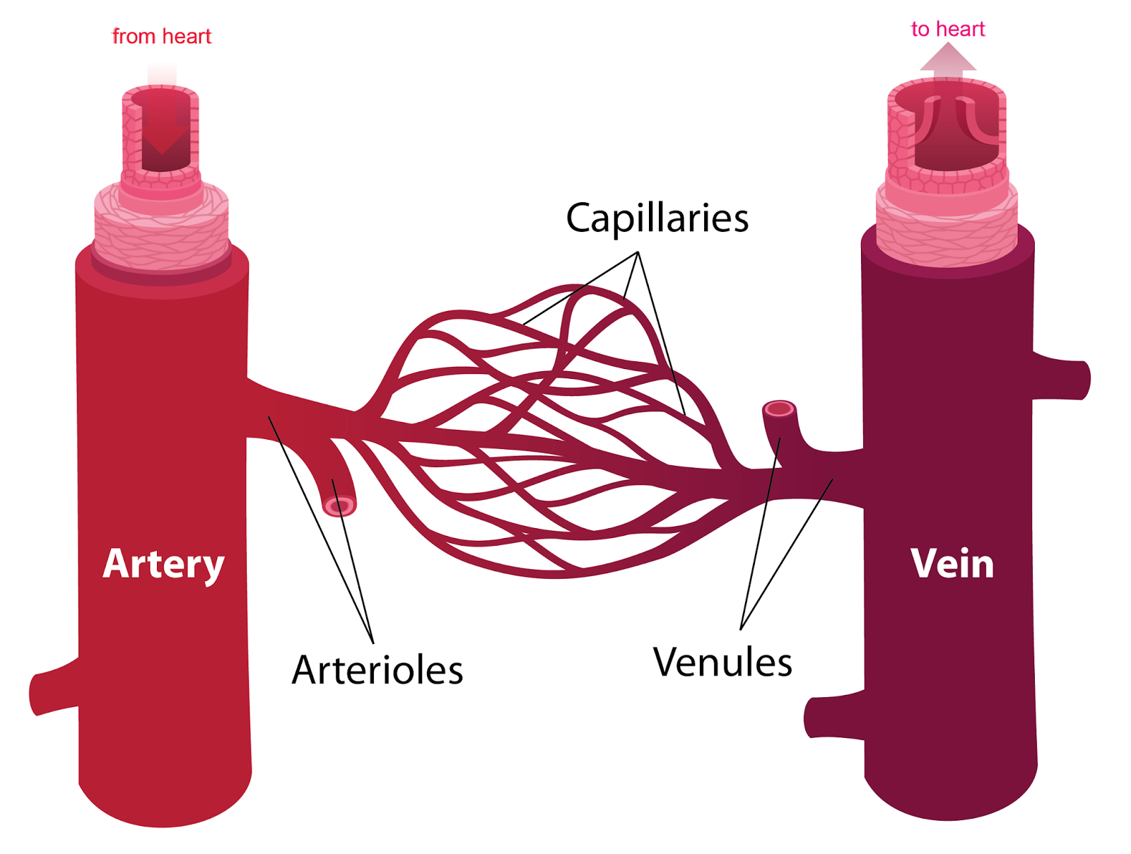
      1. Three types of blood vessels
        1. artery
        2. vein
        3. capillary
      2. Artery: Carries blood from the heart to other parts of the body.
      3. Vein: Carries blood from other parts of the body back to the heart.
      4. The arteries divides into smaller branches called arterioles.
      5. The veins receives blood from smaller branches called venules.
      6. Capillaries: Blood vessels connect the arterioles and venules.
      7. Around the capillaries are body cells.

      (Image derived from the work of Kelvinsong and is shared under the Creative Commons Attribution-Share Alike 3.0 Unported license.)


      Comparison

      Artery Vein Capillary
      Main function Carries oxygenated blood from the to ather parts of the body Carries dexoygenated blood from the other parts of the body to the heart. Connect arterioles and venules.
      Allows exchange of materials between the blood and the cells.
      Blood Carries oxygenated blood except pulmonary artery Carries deoxygenated blood except pulmonary vein
      Valves No valves except pulmonary artery All have valves except pulmonary vein No valves
      Blood pressure High Low Very low
      Blood flow Rapid Slow Very slow
      Diagram
      Wall Thick muscular wall Thin wall and less muscular Thin wall, only one-cell thick
      Size of lumen Small lumen Big lumen Very small lumen
      Branches Arterioles Venules No


      (All 3 images in the table above are derived from the work of Kelvinsong and is shared under the Creative Commons Attribution-Share Alike 3.0 Unported license.


      Recommended Videos

      Blood Vessel Structure and Function


      CIRCULATORY SYSTEM ANATOMY: Blood vessel layers model description


      1.2.5 Circulatory System in Humans

      1. The circulatory system in humans is made up of two main circulation:
        1. pulmonary circulation (between heart and lung)
        2. systemic circulation (between heart and other body parts)
      2. This is known as a double closed and complete circulatory system.

      Pulmonary Circulation

      1. Pulmonary circulation is the part of the circulation of blood in human body which carries deoxygenated blood away from the heart, to the lungs, and returns oxygenated (oxygen-rich) blood back to the heart.

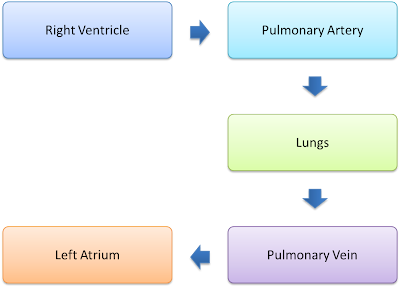
      1. Figure above shows the illustration of the pulmonary circuit.
      2. The deoxygenated blood (blue in colour) is pumped out from the right ventricle into the left and right pulmonary arteries and then to the left and right lungs.
      3. At the lungs, the deoxygenated blood receives oxygen and becomes oxygenated blood (red in colour). 
      4. The oxygenated blood is then transported from the lungs to the  pulmonary veins and then the left atrium of the heart.

      (Passage of the blood in pulmonary circulation)


      Systemic Circulation

      1. Systemic circulation is the part of the circulation of blood in human body which carries oxygenated blood away from the heart to the body, and returns deoxygenated blood back to the heart.
      2. During a systemic circulation, oxygenated blood from the left atrium of the heart is pumped into the left ventricle and then pumped out of the heart through the aorta.
      3. From aorta, the oxygenated blood is pumped to all the body tissues through arteries.
      4. Oxygen is released whereas carbon dioxide is collected from the body cells. The blood is then become deoxygenated.
      5. The deoxygenated blood is pumped back to right atrium through the vein and vena cava.

      Recommended Videos

      Circulatory system


      Other Videos

      The Circulatory System

      This video gives a complete explanation on circulatory system. However, it is a bit long.


      1.2.4 Types of Circulatory System


      Circulatory system can be classified into two types:

      1. closed circulatory system
      2. open circulatory system

      Closed Circulatory System

      1. Usually found in large animals such as vertebrates.
      2. Medium of transport is blood
      3. The blood  flows continuously in blood vessels
      4. Exchange of gases, nutrients and waste products occurs between the blood in the capillary and the body cell.

        Open Circulatory System

        1. Usually found in smaller animals such as insects and shellfish.
        2. Medium of transport is haemolymph.
        3. Haemolymph is pumped from the heart into the cavity around the body cells.
        4. Exchange of nutrients and waste products occurs directly between  and the body cells the blood around it.
        5. The haemolymph  is then pumped back to the heart.

        Close Circulatory System vs Open Circulatory System

        Open Circulatory System Close Circulatory System
        Circulating fluid Haemolymph Blood
        Occurrence Small animals such as insects Large animals such as vertebrate
        Control of fluid flow Low level of control High level of control
        Presence of valve No Yes
        Pressure of circulating fluid Relatively low Relatively high


        7.10.3 The Addition and Subtraction of Coloured Lights (Structured Questions)


        Question 1:
        Diagram 1.1 and Diagram 1.2 show an experiment to study the effect of coloured filter on white light.
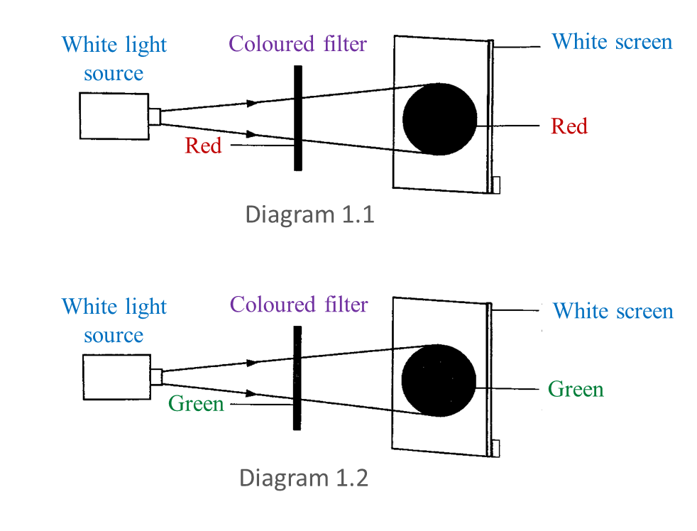

        (a) Based on this experiment, state the colour of light observed on the white screen. Write down your answer in Table below. [2 marks]


        (b) Write down one hypothesis for this experiment. [1 mark]

        (c) State one manipulated variable in this experiment. [1 mark]

        (d) Predict the colour of light observed on the white screen if blue filter is used. [1 mark]


        Answer:
        (a)

        (b)
        The colour on the screen is the same as the colour of the filter.

        (c)
        The type of colour filter.

        (d)
        Blue


        Question 2:
        Diagram 2.1 shows three coloured lights projected on a white screen.
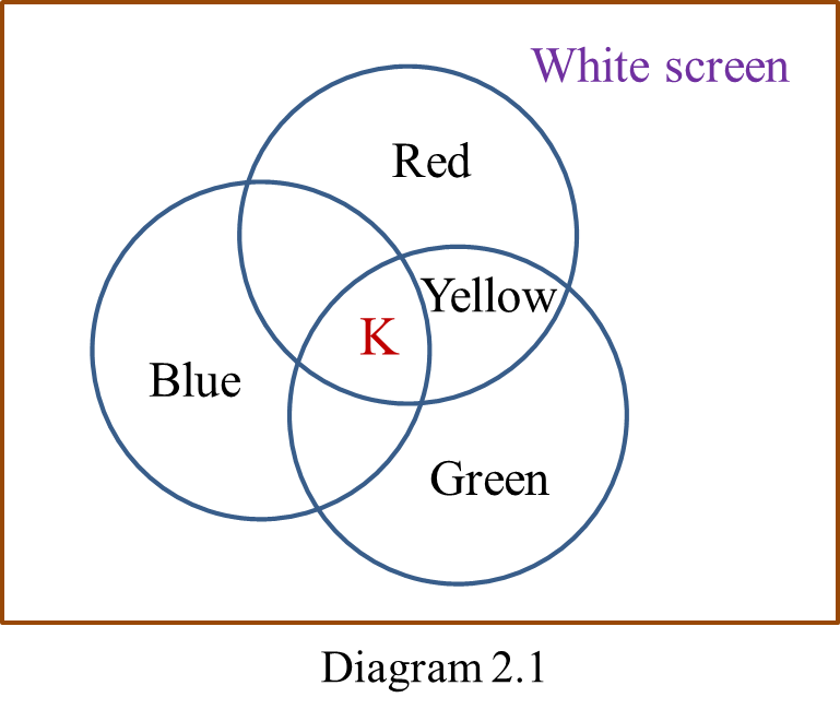


        (a) Based on the coloured lights labelled in Diagram 2.1, state
        (i) a primary colour, [1 mark]
        (ii) secondary colour. [1 mark]

        (b) What is the colour represented by K? [1 mark]

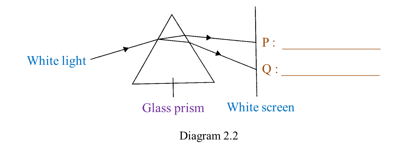
        (c) Diagram 2.2 shows a white light projected through a glass prism. [2 marks]
        In Diagram 2.2, write down the colours of lights P and Q.


        (d) A green filter is placed as shown in Diagram 2.3.
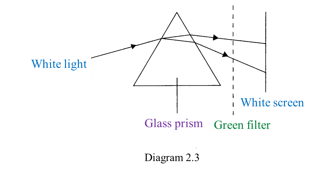
         What colour is seen on the white screen? [1 mark]


        Answer:
        (a)(i)
        Blue/ Red/ Green

        (a)(ii)
        Yellow

        (b)
        White

        (c)
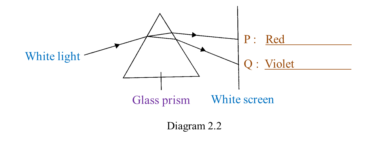
        (d) Green

        1.2.3 Function of Haemolymph in Transport


        1. Haemolymph is a fluid in the circulatory system of insects.
        2. As blood, haemolymph is the medium of transport that transport substances such as hormone and nutrient to the cells.
        3. For most insects, haemolymph is not used for oxygen transport because these animals respirate directly from their body surfaces to air.
        4. Unlike blood in human's circulatory system,the haemolymph does not flow in closed blood vessels.
        5. It is pumped out of the heart to fill the spaces between the body cells and then drawn back toward the heart through open-ended pores.

        1.2.2 Function of Blood in Transport


        Blood is an important medium in
        1. transport of oxygen and carbon dioxide
          1. In blood, oxygen combines with haemoglobin in the red blood cell to form oxyhaemoglobin.
          2. Oxygen is transported in the form of oxyhaemoglobin to the body cells which lack of oxygen.
          3. At the body cells, oxyhaemoglobin breaks down to release the oxygen to the body cells for cell respiration.
          4. Carbon dioxide is transported by the blood in ion hydrogen carbonate (ion bicarbonate) in the blood plasma carbaminohaemoglobin in the red blood cell.
        2. transport of water
        3. transport of digested food, minerals and vitamins
        4. transport of excretion
        5. transport of chemical substances such as hormones
        6. transport of heat