6.1 Types of Nutrition


[adinserter block="3"]
6.1 Types of Nutrition
 
Autotrophic Nutrition
1.  An autotroph is an organism that synthesise complex organic molecules from inorganic molecules such as air and water.
2.   Autotrophs are able to synthesise their food by
            (a)  photosynthesis
            (b)  chemosynthesis
3.  Photosynthesis is the process in which green plants and algae, called photoautotrophs, produce organic molecules from carbon dioxide and water using sunlight as a source of energy.
4.  Chemosynthesisis the process in which chemoautotrophs synthesise organic compounds by oxidizing inorganic substances such as hydrogen sulphide and ammonia.


Heterotrophic Nutrition
1.  Heterotrophs are organisms that cannot synthesise their own nutrients but instead must obtain the nutrients from other organisms.
2.  Heterotrophic nutrition is a type of nutrition in which an organism obtains energy through the intake and digestion of complex organic substances into simpler, soluble substances which are then absorbed into their bodies.
3.   Heterotrophs include animals, fungi and some bacteria.
4.   Heterotrophs may practice holozoic nutrition, saprophytism or parasitism

[adinserter block="3"]
Holozoic Nutrition
1.   Most animals like humans, herbivores and carnivores are holozoic heterotrophs.
2.   In holozoic nutrition, the organisms feed by ingesting solid organic matter which is subsequently digested and absorbed into their body.


Saprophytic Nutrition (Saprophytism)
1.   In saprophytism, the organisms called saprophytes, feed on dead and decaying organic matter.
2.   Bacteria and fungi are examples of saprophytes.
3.   Saprophytes are sometimes called decomposer.


Parasitic Nutrition (Parasitism)
1.   Parasitism is a close association in which an organism, the parasite, obtains nutrients by living on or in the body of another living organism, the host.
2.   Parasites which live on the body of the host called ectoparasitesFor examples, fleas, ticks and leeches.
3.   Parasites which live in the body of the host called endoparasites. For example, the tapeworms which infest the human intestinal tract.
 
[adinserter block="3"]

5.4.2 Meiosis (Structured Question 1 & 2)


Question 1:
Diagram I shows the different stages in a cell division.


(a)(i)
Name the type of cell division.


(a)(ii)
Arrange the stages of the cell division in the correct sequence.

(b)(i)
Explain the chromosomal behaviour in stage C.

(b)(ii)
State one importance of the chromosomal behaviour in (b)(i).

(c)
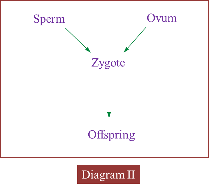
Diagram II shows the involvement of cells produced by this type of cell division in the formation of zygote.


Explain how zygote is formed.


(d)
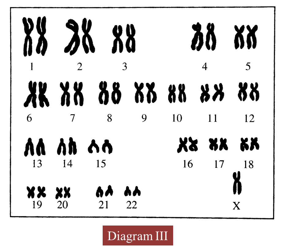
Diagram III shows the karyotype of an offspring produced.


(i)
State the number of chromosomes in the offspring.

(ii)
Name the genetic disease suffered by the offspring.

(iii)
Give one reason for the answer in (d)(ii).

(iv)
Explain how radioactive rays can cause this genetic disease.


Answer:
(a)(i)
Meiosis I

(a)(ii)

(b)(i)
The chromosomes condense, thicken and become clear.
Homologous chromosome exchange the genetic material in a process of crossing over.

(b)(ii)
Increases genetic diversity/ causes variation.


(c)
When the nucleus of a sperm (haploid) fuses with the nucleus of an ovum (haploid) during fertilization, a zygote (diploid) is formed.

(d)(i)
45/ 44 + X

(d)(ii)
Turner’s syndrome

(d)(iii)
The absence of one X chromosome which is a sex chromosome

(d)(iv)
Radioactive radiation can cause mutation of the chromosomes, as a result of which only one X chromosome is present.


5.4.1 Mitosis (Structured Question 1 & 2)


Question 1:
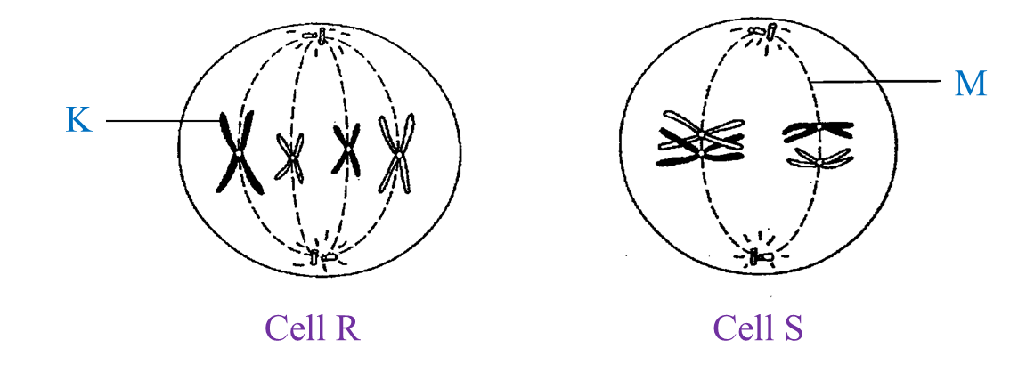
Figure I shows cells R and S undergoing cell division.


(a)(i)
Name the structures labelled K and M.

(a)(ii)
State the phase of division of cells R and S.


(b)
Cell R undergoes three consecutive divisions.

(c)(i)
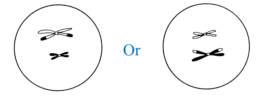
Cell S undergoes the first nuclear division and produces two cells.
Complete the diagram to show the chromosomes in one of the cells produced.

(c)(ii)
Name one organ where cell S is found.

(c)(iii)
The number of chromosomes I a somatic cell of a fly is 12.
State the number of chromosomes in a daughter cell produced at the end of the type of cell division as shown by cell S.


(d)
Cancer is a disease which causes uncontrolled growth of tissues.
Radiotherapy is a method to treat cancer by using radiation.
Explain how this treatment stops the growth of cancer cells.

(e)
An oil palm planter wants to produce a large number of oil palms in a short time.
State the best technique to be used by the planter and one problem to be considered in using the technique.


Answer:
(a)(i)

K: Chromatid
M: Spindle fibre

(a)(ii)
Cell R: Metaphase
Cell S: Metaphase I

(b)
8 daughter cells

(c)(i)


(c)(ii)
1. Testis
2. Ovary

(c)(iii)
6 chromosomes

(d)
Radioactive rays destroy the cells, so the rate of mitosis is controlled.

(e)
- Cloning / culture tissue
- All the clones are easily attacked by diseases because all the clones have the same resistance towards diseases.


4.7.2 Enzymes (Structured Question 1 & 2)


Question 1:
Diagram I shows the organelles involved during the synthesis and secretion of an enzyme in an animal cell.

(a)
Name the organelles labelled J and L.

(b)(i)
State the function of organelle M.
(b)(ii) Explain the role of organelle K in the synthesis of the enzyme.


(c)
Enzymes are widely used in our daily life and industries.
Explain how enzymes act in:
(i) helping to cook meat.
(ii) extracting agar from seaweeds.


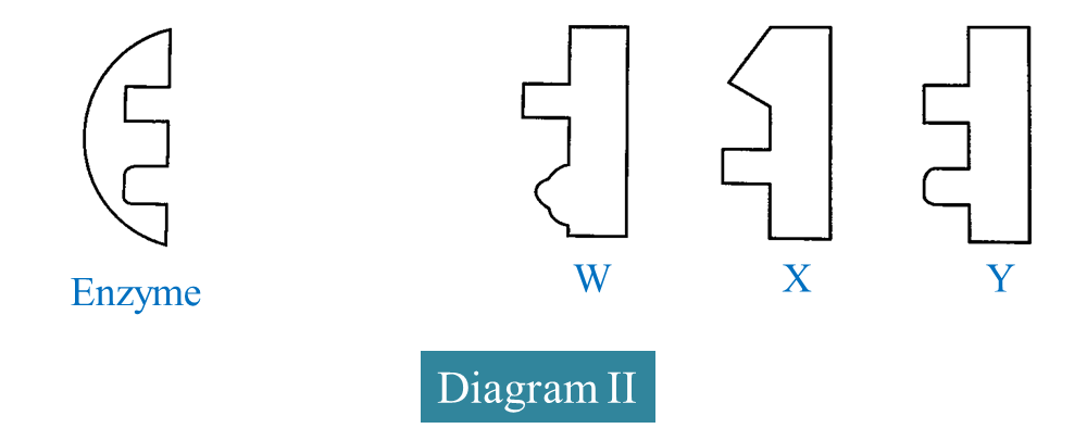
(d) Diagram II shows the structure of an enzyme and three substrates W, X and Y.


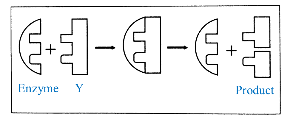
Based on Diagram II, complete the schematic diagram below to show the mechanism of enzyme action on a suitable substrate.


Answer:
(a)
J: Golgi apparatus
L: Secretory vesicles

(b)(i)
Produces energy

(b)(ii)
K stores genetic information in the DNA and this information is transferred to the RNA which then carries it out to the cytoplasm.


(c)(i)
Protease enzyme softens the meat.

(c)(ii)
Cellulase breaks down cell walls of seaweed and frees agar contained in it.

(d)


4.7.1 Proteins (Structured Question 1 & 2)


Question 1:
Diagram I shows two levels of organization in protein structure.


(a) In Diagram I, name the level of organization in the protein structures of P and Q.


(b)
Describe the structure of protein P.

(c)
Diagram II shows how protein P is broken down to form products.


(d) Explain how the products in Diagram II are formed.


(e)
State why animal proteins are first class protein.

Silk is an example of protein. A silk garment crumples when it is washed using hot water at a temperature of 65OC.
(i) Explain the above statement.

(ii)
Based on the above statement, suggest two ways to maintain the quality of silk garments.


Answer:
(a)
P: Primary structure
Q: Secondary structure

(b)
The linear sequence of amino acids in a polypeptide chain.

(c)
Protein P is broken down by an enzyme (polypeptidase) in hydrolysis to form dipeptides.

(d)
Because it contains all the 8 essential amino acids needed by humans. 


(e)(i)
Silk is a protein fibre. High temperatures will reduce the strength of the hydrogen bonds and disrupt the structure, thus denaturation of the protein occurs.

(e)(ii)
1.Wash it in cold water
2. Do not wring
3. Do not expose to direct sunlight
4. Never use chlorine bleach on silk
(Choose any 2)

4.2.2 Disaccharides


  1. Disaccharides are formed when two monosaccharides combine together through a chemical process called the condensation.
  2. Disaccharides are known as complex sugar.
  3. Examples of disaccharides are,
    1. Maltose 
      1. known as malt sugar
      2. It is a reducing sugar
      3. It is formed from condensation of two glucose molecules.
        Glucose + glucose  maltose + water 
      4. Found in germinating cereals
      5. Produced during digestion of starch
    2. Sucrose
      1. It is found in sugar cane, beet root and  sweet fruits
      2. It is a non-reducing sugar
      3. It is  formed from glucose and fructose.
        Glucose + fructose → sucrose + water
    3. Lactose
      1. Present in all mammals
      2. Is a reducing sugar
      3. It is formed from glucose and galactose
        Glucose + galactose  lactose + water
  4. Disaccharides are decomposed to units of monosaccharide through hydrolysis (the addition of one molecule of water)

4.2.1 Monosaccharides

  1. Monosacharides are monomers of carbohydrates. 
  2. Examples of monosacharides are:
    1. Glucose — found in plants and animals (It is the energy source for cellular respiration)
    2. Fructose — found in fruits and honey
    3. Galactose — present in milk
  3. They are reducing sugars, they can remove oxygen from other compounds..
  4. They can be detected by using Fehling's or Benedict's test. 
  5. When monosaccharides are heated with Fehling's or Benedict's solution, they will reduce the blue copper(II) sulphate in the two solutions to a brick red precipitate.

4.2 Carbohydrate


  1. Carbohydrate consist of the elements carbon, hydrogen and oxygen. 
  2. The H : O ratio in all carbohydrate molecules is 2 : 1.
  3. Examples of carbohydrate include starch, sugar, glycogen and cellulose.
  4. Excess carbohydrates are stored in the form of glycogen in the liver. Some are converted to fat and stored in the adipose tissues below the skin and around organs.
  5. There are three types of carbohydrates:
    1. Monosaccharides
    2. Disaccharides
    3. Polysaccharides

4.1 Chemical Composition of the Cell


  1. The elements found in the cell include
    1. carbon, 
    2. oxygen, 
    3. hydrogen, 
    4. nitrogen, 
    5. sulphur, 
    6. phosphorus, 
    7. calcium,
    8. sodium,
    9. potassium, 
    10. magnesium,
    11. iron 
    12. chlorine, 
  2. The chemical compounds in the cell can be classified into two groups, 
    1. organic: compounds which contain the element carbon and originate from living things. Example: carbohydrates, lipids, proteins and nucleic acids
    2. inorganic: compounds which do not originate from living things. Example: water and mineral salts.
  3. .Organic compounds are synthesised by the cells themselves whereas inorganic compounds are not synthesised by the cells themselves but are obtained from the external environment.